
Title
Subtitle
Price
Under eye filler migration can be managed safely. Discover expert prevention tips, correction techniques, and patient care strategies to improve outcomes.

The delicate under-eye area is one of the most challenging yet in-demand regions for dermal filler treatment. However, it also carries some of the highest risks for aesthetic complications. Therefore, understanding under-eye filler migration is critical to optimizing results and patient safety.
With growing demand for non-surgical tear trough correction, training has become essential. Explore aesthetics courses for doctors, especially tear trough filler training offered by the HubMed Ed learning platform, to build anatomical confidence and clinical precision.
With proper technique and product choice, under-eye fillers, especially HA-based ones-can be both effective and safe. However, the anatomical complexity of this area demands a skilled hand and thoughtful planning.
While hyaluronic acid fillers offer the advantage of reversibility with hyaluronidase, their success depends greatly on injector experience, appropriate patient selection, and conservative dosing. Even small errors in this area can result in visible complications or undesirable outcomes. Therefore, “safe” should not be equated with “risk-free.” Practitioners must assess each case individually and use precise, conservative injection techniques.
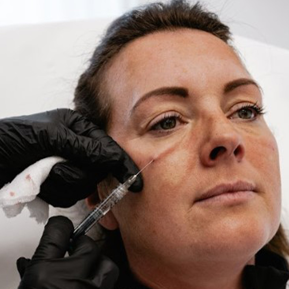
Thin skin, limited subcutaneous fat, poor lymphatic drainage, and proximity to key vascular structures increase the likelihood of complications in the under-eye region. Patients often seek treatment for multiple overlapping concerns, such as skin laxity, dark circles, and hollowness-making precision essential.
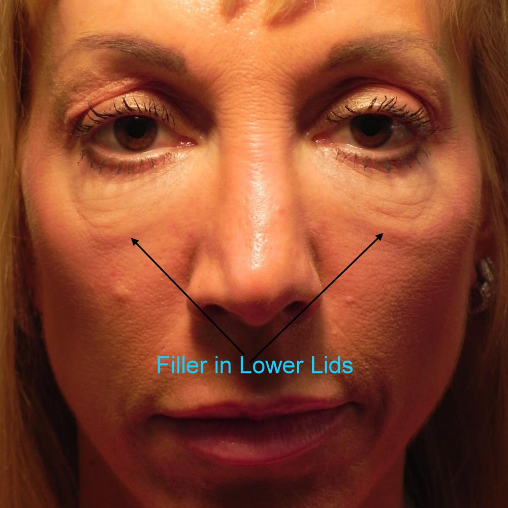
Even minor misplacement of the product can lead to issues like puffiness or the Tyndall effect, underscoring the need for advanced anatomical understanding. Without proper anatomical knowledge and product selection, tear trough filler complications such as puffiness, discoloration, and migration can occur even with minimal product volume.
There are several contributing factors to under-eye fillers gone wrong, such as migration, many of which relate to poor technique, unsuitable product, or individual anatomy. These include:
Other contributing factors include repetitive facial movements, lymphatic insufficiency, and patient-specific anatomy such as malar edema or festoons. These increase the likelihood of migrated under-eye filler and visible swelling over time.
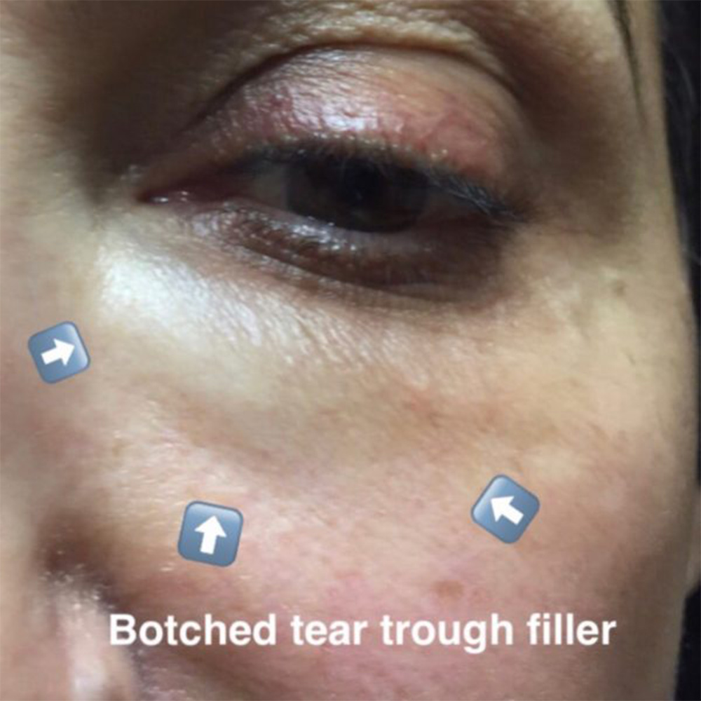
Migrated under eye filler may not appear immediately. Practitioners should differentiate between early post-treatment edema and true migration. Key signs include persistent or delayed puffiness, blue-gray discoloration (Tyndall effect), palpable lumps, or asymmetry.
A thorough assessment includes patient history (product, volume, technique, timeline) and physical exam. In complex cases, diagnostic ultrasound can aid in visualizing filler location and guiding correction. Don’t mistake festoons or fat pad prolapse for filler-related issues, they often mimic migrated filler under eyes.
Practitioners often ask how to fix under-eye filler issues, particularly in cases of migration or puffiness. When managing under-eye filler complications, hyaluronidase remains the primary corrective option for HA-based products due to its precision and reversibility. Proper dilution, depth, and conservative volume are key to targeted reversal without causing excessive tissue disruption.
Tyndall effect correction typically involves the targeted use of hyaluronidase to dissolve superficially placed filler. Hyaluronidase for under-eye correction should be used with caution, balancing effective resolution with the risk of tissue disruption.
In less severe cases, massage or time may allow the filler to settle or resolve. For stubborn nodules or excessive product, aspiration may be useful. In cases where filler is not suitable due to tissue quality, PRF EZ Gel or other regenerative therapies may offer a safer, biologically active solution.
The most effective strategy for managing under-eye filler complications is prevention. Practitioners can minimize risks by following these clinical best practices:
Use blunt cannulas when appropriate to minimize trauma and vascular penetration. For high-risk cases, additional education is key. Consider the Aesthetics Complications Masterclass Course at HubMed Ed to develop advanced complication management skills.
Not every patient is an ideal candidate for under-eye filler, and proper screening can make the difference between a smooth result and a complicated correction. Contraindications include chronic edema, poor skin tone, festoons, malar bags, and a history of previous complications.
Take a detailed history of all prior aesthetic procedures, systemic conditions like thyroid dysfunction, and use of medications affecting fluid balance. Also, emphasize that tear trough correction may not be a suitable procedure for some patients, and that alternatives may be preferable.
Incorporating high-frequency ultrasound has reshaped how practitioners approach under-eye filler treatments, offering real-time visual guidance and improved diagnostic accuracy. It enables clinicians to:
Many advanced aesthetic practices now consider ultrasound guidance an integral part of safe and effective filler procedures. They help minimize risk, avoid trial-and-error dissolving, and improve patient safety.
Clear and proactive communication helps minimize anxiety and ensures patients are well-prepared throughout the treatment process. Begin by setting realistic expectations about what under-eye filler can and cannot achieve, including the expected duration of results. Explain the possibility of side effects, like minor swelling or bruising, post-treatment.
Provide written aftercare instructions, including avoiding high-sodium foods, strenuous exercise, massaging the area, and alcohol for 24-48 hours. Follow-up appointments are essential to assess symmetry and early signs of complications. Educated patients are less likely to panic over normal healing responses and more likely to report abnormal changes promptly.
With careful planning, advanced technique, and ongoing education, migrated filler doesn’t have to derail outcomes. Stay current, stay conservative, and stay curious!
For deeper insight, explore our hyaluronidase tutorials, complication management modules, and case-based injector training designed for real-world success.
In some patients, minor filler migration may resolve over time as the product is naturally metabolized. However, persistent or aesthetically concerning cases often require targeted dissolution with hyaluronidase.
Swelling typically occurs within the first few days post-injection and gradually subsides. Migration, on the other hand, appears later and may involve contour irregularities, puffiness, or bluish discoloration.
Low-density, low-hydrophilic hyaluronic acid fillers formulated for tear trough treatments are least likely to migrate. These products integrate more smoothly and exert less tissue pressure, reducing displacement risk.
Juvéderm Voluma is a high G’ filler intended for midface volumization and not for the delicate under-eye area. When used improperly in this region, it carries a greater risk of migration and visible lumpiness.
Yes, particularly if injected too medially or in areas with poor anatomical support. Excess product or soft tissue mobility can allow filler to drift toward the tear trough over time.
This article is intended for licensed medical professionals. All protocols, dosages, and treatment insights referenced herein are based on published literature. The content is not intended to encourage application, diagnosis, or self-treatment of unlicensed individuals, and should not be used as a substitute for the clinical judgment of a qualified healthcare provider.
Under eye filler migration can be managed safely. Discover expert prevention tips, correction techniques, and patient care strategies to improve outcomes.
The delicate under-eye area is one of the most challenging yet in-demand regions for dermal filler treatment. However, it also carries some of the highest risks for aesthetic complications. Therefore, understanding under-eye filler migration is critical to optimizing results and patient safety.
With growing demand for non-surgical tear trough correction, training has become essential. Explore aesthetics courses for doctors, especially tear trough filler training offered by the HubMed Ed learning platform, to build anatomical confidence and clinical precision.
With proper technique and product choice, under-eye fillers, especially HA-based ones-can be both effective and safe. However, the anatomical complexity of this area demands a skilled hand and thoughtful planning.
While hyaluronic acid fillers offer the advantage of reversibility with hyaluronidase, their success depends greatly on injector experience, appropriate patient selection, and conservative dosing. Even small errors in this area can result in visible complications or undesirable outcomes. Therefore, “safe” should not be equated with “risk-free.” Practitioners must assess each case individually and use precise, conservative injection techniques.
Thin skin, limited subcutaneous fat, poor lymphatic drainage, and proximity to key vascular structures increase the likelihood of complications in the under-eye region. Patients often seek treatment for multiple overlapping concerns, such as skin laxity, dark circles, and hollowness-making precision essential.
Even minor misplacement of the product can lead to issues like puffiness or the Tyndall effect, underscoring the need for advanced anatomical understanding. Without proper anatomical knowledge and product selection, tear trough filler complications such as puffiness, discoloration, and migration can occur even with minimal product volume.
There are several contributing factors to under-eye fillers gone wrong, such as migration, many of which relate to poor technique, unsuitable product, or individual anatomy. These include:
Other contributing factors include repetitive facial movements, lymphatic insufficiency, and patient-specific anatomy such as malar edema or festoons. These increase the likelihood of migrated under-eye filler and visible swelling over time.
Migrated under eye filler may not appear immediately. Practitioners should differentiate between early post-treatment edema and true migration. Key signs include persistent or delayed puffiness, blue-gray discoloration (Tyndall effect), palpable lumps, or asymmetry.
A thorough assessment includes patient history (product, volume, technique, timeline) and physical exam. In complex cases, diagnostic ultrasound can aid in visualizing filler location and guiding correction. Don’t mistake festoons or fat pad prolapse for filler-related issues, they often mimic migrated filler under eyes.
Practitioners often ask how to fix under-eye filler issues, particularly in cases of migration or puffiness. When managing under-eye filler complications, hyaluronidase remains the primary corrective option for HA-based products due to its precision and reversibility. Proper dilution, depth, and conservative volume are key to targeted reversal without causing excessive tissue disruption.
Tyndall effect correction typically involves the targeted use of hyaluronidase to dissolve superficially placed filler. Hyaluronidase for under-eye correction should be used with caution, balancing effective resolution with the risk of tissue disruption.
In less severe cases, massage or time may allow the filler to settle or resolve. For stubborn nodules or excessive product, aspiration may be useful. In cases where filler is not suitable due to tissue quality, PRF EZ Gel or other regenerative therapies may offer a safer, biologically active solution.
The most effective strategy for managing under-eye filler complications is prevention. Practitioners can minimize risks by following these clinical best practices:
Use blunt cannulas when appropriate to minimize trauma and vascular penetration. For high-risk cases, additional education is key. Consider the Aesthetics Complications Masterclass Course at HubMed Ed to develop advanced complication management skills.
Not every patient is an ideal candidate for under-eye filler, and proper screening can make the difference between a smooth result and a complicated correction. Contraindications include chronic edema, poor skin tone, festoons, malar bags, and a history of previous complications.
Take a detailed history of all prior aesthetic procedures, systemic conditions like thyroid dysfunction, and use of medications affecting fluid balance. Also, emphasize that tear trough correction may not be a suitable procedure for some patients, and that alternatives may be preferable.
Incorporating high-frequency ultrasound has reshaped how practitioners approach under-eye filler treatments, offering real-time visual guidance and improved diagnostic accuracy. It enables clinicians to:
Many advanced aesthetic practices now consider ultrasound guidance an integral part of safe and effective filler procedures. They help minimize risk, avoid trial-and-error dissolving, and improve patient safety.
Clear and proactive communication helps minimize anxiety and ensures patients are well-prepared throughout the treatment process. Begin by setting realistic expectations about what under-eye filler can and cannot achieve, including the expected duration of results. Explain the possibility of side effects, like minor swelling or bruising, post-treatment.
Provide written aftercare instructions, including avoiding high-sodium foods, strenuous exercise, massaging the area, and alcohol for 24-48 hours. Follow-up appointments are essential to assess symmetry and early signs of complications. Educated patients are less likely to panic over normal healing responses and more likely to report abnormal changes promptly.
With careful planning, advanced technique, and ongoing education, migrated filler doesn’t have to derail outcomes. Stay current, stay conservative, and stay curious!
For deeper insight, explore our hyaluronidase tutorials, complication management modules, and case-based injector training designed for real-world success.
In some patients, minor filler migration may resolve over time as the product is naturally metabolized. However, persistent or aesthetically concerning cases often require targeted dissolution with hyaluronidase.
Swelling typically occurs within the first few days post-injection and gradually subsides. Migration, on the other hand, appears later and may involve contour irregularities, puffiness, or bluish discoloration.
Low-density, low-hydrophilic hyaluronic acid fillers formulated for tear trough treatments are least likely to migrate. These products integrate more smoothly and exert less tissue pressure, reducing displacement risk.
Juvéderm Voluma is a high G’ filler intended for midface volumization and not for the delicate under-eye area. When used improperly in this region, it carries a greater risk of migration and visible lumpiness.
Yes, particularly if injected too medially or in areas with poor anatomical support. Excess product or soft tissue mobility can allow filler to drift toward the tear trough over time.
This article is intended for licensed medical professionals. All protocols, dosages, and treatment insights referenced herein are based on published literature. The content is not intended to encourage application, diagnosis, or self-treatment of unlicensed individuals, and should not be used as a substitute for the clinical judgment of a qualified healthcare provider.