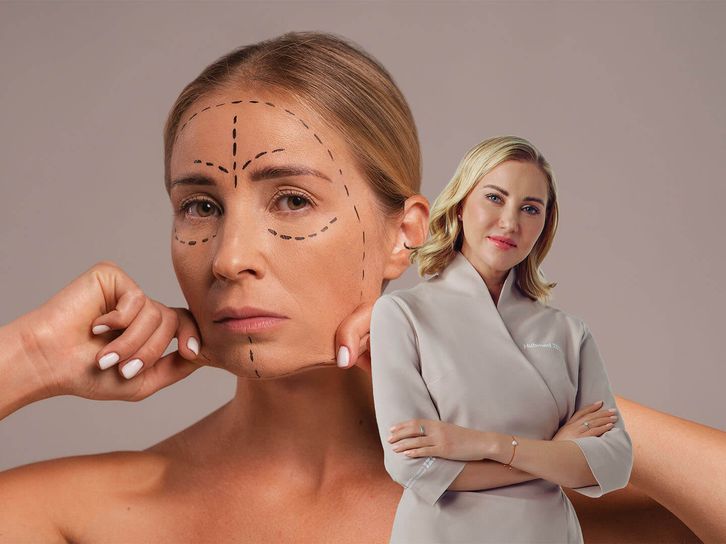
Introduction:
Amidst a surge in popularity and a concerning lack of regulation, safeguarding patient well-being while administering injectables is essential. While achieving a desired aesthetic outcome is certainly a goal, the foundation of this practice rests firmly on a deep understanding of facial anatomy. Beneath every face lies a complex network of muscles, nerves, blood vessels, and soft tissues. With a better understanding of facial anatomy, injectors can:
- Strategically place injectables to achieve balanced and natural-looking outcomes.
- Emphasizing a safe and controlled approach can reduce complications. This minimizes the risk of bleeding, bruising, or nerve damage.
- Assess individual variations in facial structure and tailor treatment plans accordingly.
- Educate their patients, explain different treatment options, potential risks, and benefits, and empower them to make informed decisions about their care. This ultimately cultivates a trusting patient-practitioner relationship.
The art of understanding facial anatomy has moved beyond the limitations of a 2-dimensional illustration. We've moved beyond flat pictures to a 3D understanding, allowing for safer and more effective injections. This includes navigating the 5 facial layers and newly discovered structures, all while avoiding vascular danger zones. Finally, the field of facial anatomy is constantly evolving, as it is reflected in the ongoing development and refinement of its terminology. This ensures clear communication within the field and fosters continuous advancements in safe and effective injectable practices.
Age-Related Changes:
Facial aging is a well-documented phenomenon characterized by interrelated changes within the facial structures.1 This progressive process involves the skeletal system, musculature, adipose tissue, and the integumentary system. For example, age-related bone resorption, particularly in the mandible, can result in a reduction in structural support for overlying soft tissues. Additionally, facial musculature undergoes progressive atrophy, which reduces its ability to keep skin firm and create expressive movements.
Facial contours too, witness a change. The distribution of adipose tissue undergoes a dynamic shift, with lipoatrophy (volume loss) occurring in the malar region (cheeks) and lipohypertrophy (accumulation) in the submental region. The supportive framework weakens with age due to a decline in the synthesis of collagen and elastin. This intrinsic aging process contributes to the development of rhytids (wrinkles), cutis laxa (loss of skin firmness), and a decline in overall skin quality.
These changes show up in different ways:
- Volume depletion, a consequence of reduced bone density, fat pad migration, and muscle atrophy, contributes to a hollowed appearance, particularly in the midfacial and temporal regions.
- Soft tissue ptosis (descent) occurs due to weakened musculature and decreased skin elasticity, resulting in visible sagging and the formation of jowls.
- Dynamic imbalance arising from altered muscle function. This disrupts the delicate balance between facial expressions and leads to the formation of persistent rhytids (static wrinkles) and a loss of overall facial harmony.
For more clarity, the following tables detail the effects of aging on different tissues within the upper, middle, and lower thirds of the face.
Understanding the underlying anatomical and physiological basis of facial aging allows to develop precise interventionsfor patients, including,
- Volume restoration with dermal fillers to address facial hollowness and re-establish a more youthful contour.
- Skin rejuvenation techniques such as laser therapy or microneedling to stimulate collagen production, improve skin texture and elasticity.
- Muscle rejuvenation with botulinum toxin injections (Botox) to treat dynamic wrinkles caused by hyperactive muscles.
Interestingly, the rate and severity of these changes can vary with individual factors such as genetics, ultraviolet radiation exposure (sun exposure), and lifestyle habits.
The Skin
The skin, our body's largest and most visible organ, shields us from the external environment while simultaneously contributing to our aesthetic appeal.
The Epidermis:
The outermost layer, the epidermis, is a dynamic and cell-rich environment. Keratinocytes, the predominant cell type, undergo a continuous differentiation process, maturing to form the skin's protective barrier. Scattered amongst these "brick and mortar" cells are specialized pigment-producing melanocytes. These cells determine our skin color variation and offer sun protection through melanin production. Additionally, Langerhans cells, acting as vigilant sentries, play a crucial role in the skin's immune function by presenting antigens to the body's defense system.
The Dermis:
Beneath the epidermis lies the dermis, the structural foundation of the skin. This layer is considered the outermost layer of the superficial fascia. Here, a network of proteins and other substances, known as the extracellular matrix, provides strength and support. Type I collagen makes up the most abundant protein in this network, offering remarkable tensile strength. Other collagen types, elastin, proteoglycans, and fibronectins also contribute to the dermis' structural integrity and functional properties. The dermis has a rich vascular plexus that carries oxygen and nutrients to the skin.
Thickness and Function: A Delicate Balance
The thickness of the dermis is not uniform across the face. It strategically adapts to different functional needs. In areas requiring greater structural support, such as the forehead and nasal tip, the dermis is thicker. Conversely, in areas with enhanced mobility, like the eyelids, the dermis is thinner. However, this delicate balance between thickness and function has implications for aging. Thinner regions may be more prone to the visible signs of aging, highlighting the importance of maintaining skin health and integrity throughout life.
Collagen imparts resilience, tensile strength, adherence, and thickness to the skin, while elastin enhances its elasticity. Hyaluronic acid (GAG) ensures optimal hydration, essential for sustaining skin quality, including its natural glow, luminosity, and overall health.
Subcutaneous Layer
Understanding the architecture and composition of the subcutaneous layer, or the hypodermis is essential for navigating facial procedures effectively. This layer comprises two key components with distinct functionalities:
- Subcutaneous Adipose Tissue (Subcutaneous Fat): This primary component is composed of adipocytes (fat cells), and acts as a volume sculptor, dictating facial contours and providing essential volume.The distribution and arrangement of this fat exhibit significant regional variation across the face. In areas like the scalp, the subcutaneous layer boasts a uniform thickness and consistent connection to the overlying dermis.
However, the facial landscape is far more diverse, with specialized regions such as the eyelids and lips demonstrating a remarkably compact subcutaneous layer, often appearing devoid of significant adipose tissue. Conversely, regions like the nasolabial folds exhibit a considerably thicker layer. Interestingly, areas with abundant subcutaneous tissue tend to show greater susceptibility to the effects of aging, as the anchoring retinacular cutis fibers may weaken and distend over time.
- Fibrous Retinacular Cutis: This intricate network of collagen fibers acts as a bridge that binds the dermis (the skin's inner layer) to the underlying SMAS (Superficial Musculoaponeurotic System). The retinacular cutis is a specialized portion of the retaining ligaments that extends through the subcutaneous tissue. Its attachment pattern is particularly noteworthy - a stronger and denser connection is formed with the dermis compared to the SMAS. The retinacular cutis fibers function similarly to a tree with its roots anchoring it firmly to the ground. As they rise through the SMAS, these initially thicker fibers progressively branch out into finer microligaments that connect to the dermis. This explains why deeper subcutaneous dissection, closer to the SMAS, is often easier. Here, fewer retinacular cutis fibers are present, and the adipose tissue doesn't directly attach to the SMAS surface.
Furthermore, the retinacular cutis isn't uniform across the face. Fiber orientation and density vary depending on the underlying structures. Vertically oriented fibers are most concentrated in areas corresponding to retaining ligaments, providing superior support and compartmentalizing the subcutaneous fat.
These zones, such as McGregor's patch over the zygoma, often necessitate a sharper dissection technique for mobilization. In contrast, areas between these retaining ligaments, known as soft tissue spaces, exhibit less dense, more horizontally oriented retinacular fibers. Consequently, these spaces allow for easier mobility of the superficial facial layers over the deeper structures, often requiring only blunt finger dissection.
Facial Adipose Compartments
The facial adipose tissue (fat) compartments are categorized as superficial and deep. Let’s explore their location, structure, and influence on facial aesthetics.
Superficial vs. Deep: A Tale of Two Compartments
Facial fat is not a homogenous mass but a collection of distinct compartments classified based on their depth relative to the superficial musculoaponeurotic system (SMAS).
Superficial Fat Compartments:
These reside above the SMAS and exhibit a more mobile nature. They are further categorized:
- Forehead: Three compartments exist—central, middle, and lateral temporal cheek. The middle temporal compartment flanks the central compartment, and the lateral temporal cheek compartment connects to the lateral cheek and cervical fat.
- Midface: This region includes medial, middle, and lateral temporal cheek compartments alongside the nasolabial fat pad.
- Lower Face: Jowl fat forms the superficial layer here, along with mental and submental fat. Interestingly, jowl fat occupies a specific location - medial to lateral temporal cheek fat, lateral to nasolabial fat, and superior to the mandible.
- Periorbital Region: Three superficial compartments are present - superior, inferior, and lateral orbital fat.
Structural Characteristics of Superficial Fat
Superficial fat compartments are characterized by:
- Small, Tightly Packed Lobules: The fat lobules within these compartments are relatively small and arranged in a continuous, uniform manner.
- Mobility: Due to their location above the SMAS, they exhibit greater mobility and are influenced by both resting and dynamic facial expressions mediated by mimetic muscles.
Deep Fat Compartments: Anchors and Support
Lying beneath the SMAS are the deep fat compartments. These offer structural support and differ from their superficial counterparts in several ways:
- Location: Deep fat compartments are firmly anchored to the underlying bone.
- Lobules: These compartments house larger, more loosely arranged fat lobules, often with a less organized pattern.
- Function: Deep fat provides essential functions:some text
- Contouring: It contributes to the overall facial shape and definition.
- Support: It provides structural support for the overlying superficial fat compartments.
- Gliding Plane: It facilitates smooth muscle movement by acting as a gliding plane.
- Immobility: Unlike superficial fat, deep fat exhibits minimal movement due to its firm anchorage to the bone.
Examples of Deep Fat Compartments:
- Deep medial cheek fat
- Buccal fat
- Medial and lateral suborbicularis oculi fat (SOOF)
- Retro-orbicularis oculi fat
Functional Significance and Aging:
- The deep fat layer plays a vital role in maintaining facial fullness and youthful contours.
- With age, this layer may undergo disintegration and descent. This can lead to a more prominent lower eyelid border, emphasizing the malar crescent and nasojugal fold (smile lines).
- Hormonal changes, particularly post-menopausal estrogen decline, can further impact this layer. While superficial fat may decrease, the deep layer may experience increased fat deposition that contributes to facial volume shifts.
The Superficial Musculoaponeurotic System (SMAS)
The Superficial Musculoaponeurotic System (SMAS) forms a distinct anatomical environment for facial expression muscles, differentiating them from deeper skeletal muscles beneath the fascia. Unlike their counterparts, facial muscles reside within the superficial fascia, allowing them to move the overlying soft tissues directly.
Nearly all these muscles of expression have a significant portion, if not their entire course, within the SMAS, concentrated around the orbital and oral cavities for optimal function. The occipitofrontalis muscle, responsible for scalp movement, is an exception and lies superficial to the SMAS.
Interestingly, the SMAS maintains continuity across the face with regional variations in terminology based on the underlying muscle: galea aponeurosis on the scalp, temporoparietal fascia on the temple, orbicularis fascia around the eye, and platysma in the neck.
Within the SMAS itself, a layered organization exists. Broad, flat muscles form the superficial layer, primarily covering the anterior aspect of the face with minimal bony attachments. These muscles rely on vertically oriented retaining ligaments for stability. Deeper within the SMAS are muscles responsible for more precise control of facial expressions, such as frowning, smiling, lip elevation and depression, and chin movement.
Importantly, the SMAS, along with the overlying subcutaneous fat and skin, forms a distinct mobile unit that is separate from the deeper, fixed facial structures. Specialized ligaments known as retinacula cutis connect the mobile unit to these deeper structures.
With aging, these ligaments weaken and compromise their support for the mobile unit. This loss of support allows the unit to shift position relative to the underlying fixed structures. This in turn, contributes to the characteristic facial changes we observe in aging, such as fat descent and a "rounder" appearance.
The Sub-SMAS Layer
This stratum is crucial for sub-SMAS facelifts, as it harbors a complex interplay of structures with distinct functionalities:
- Soft Tissue Spaces: These spaces, acting as musculofascial gliding planes, facilitate independent movement of the peri-orbital and peri-oral facial expression musculature. These muscles can move freely over the deeper fascia responsible for mastication directly beneath them.
- Retaining Ligaments: Strategically positioned at the borders of the soft tissue spaces, these ligaments function as reinforcing elements to maintain the integrity and definition of these spaces.
- Deep Muscle Layers: This layer also serves as a transitional zone for the deeper layers of facial expression musculature. Here, these muscles transition from their bony attachments to their more superficial origins within the soft tissues.
- Facial Nerve Branches: These branches course through layer 4, traversing from deep to superficial regions to innervate their target muscles of facial expression.
Functional Significance and Implications:
The existence of soft tissue spaces within this layer allows for independent movement of the overlying mobile facial expression muscles over the deeper, more stable fascia used for chewing.2-4,6
Retaining ligaments play a vital role in maintaining the boundaries of these spaces and providing additional support. In areas like the lateral face near the ear, where facial movement is minimal, the soft tissue space disappears. Layers 1-5 (skin to parotid gland capsule) fuse together, forming a robust retaining ligament area ideal for secure surgical fixation with sutures (e.g., platysma auricular fascia).
Conversely, the anterior face, with its highly mobile areas around the eyes and mouth, exhibits a distinct arrangement. Here, the retaining ligaments become more compact and strategically positioned around the bony cavities, providing the final point of deep fascial support for the mobile eyelids and lips. Additionally, these ligaments also serve as crucial transition points for facial nerve branches as they travel from deeper regions to innervate specific muscles.
Retaining Ligament
Facial retaining ligaments, a network of supportive structures, significantly impact wrinkle formation and facial aesthetics. These ligaments provide a framework by connecting facial tissues like skin and muscle to the underlying bone. Two main types exist:5
- True ligaments: These run directly from bone to skin, offering strong support. An example is the maxillary ligament, which influences the nasolabial fold.
- False ligaments: These originate from deeper tissues like muscles or fat pads. The buccal part of the bucomaxillary ligament is an example, providing less structural support.
The influence of ligaments on wrinkles varies across the face. Some key connections existing in the midface include:
- Midcheek crease: This wrinkle is closely associated with the zygomatic ligament.
- Labiomandibular fold (marionette line): The mandibular ligament plays a role in its formation.
- Tear trough: This deformity is influenced by the tear trough ligament, part of the orbital retaining ligament complex.
The cause of the nasolabial fold remains a topic of debate. The fascial theory suggests retaining ligaments like the maxillary ligament are the primary culprit. However, the muscular theory, which attributes it to muscles like the zygomaticus and levator labii superioris, currently holds slightly more weight based on recent research.
Beyond wrinkles, ligaments also play a part in jowl formation. The labiomandibular fold and other ligaments, like the mandibular septum and temporal septi help prevent jowls by separating fat compartments and defining facial spaces. The masseteric cutaneous ligament's role is more intricate. It provides a reference point for facial boundaries and may influence jowl formation by affecting how the buccal fat pad protrudes.
Facial Spaces
Understanding the complex network of soft tissue spaces within the subSMAS layer is essential in cosmetic facial procedures. These spaces, meticulously defined by their boundaries and fortified by retaining ligaments, serve as anatomically "safe zones" for surgical dissection. They remain devoid of vital structures and facial nerve branches and minimize the risk of inadvertent damage. However, the roof of each space, being the least supported, is susceptible to laxity over time, which contributes significantly to age-related facial changes.4
Clear identification of these areas allows for specific release of retaining ligaments to aid in desired movement while safeguarding essential structures. Here is a concise overview of the surgically significant facial soft tissue spaces.
Deep Fascia
The deepest layer of the facial soft tissue isn't a uniform sheet but rather an adaptation to the structures it supports. Where the face meets bone, the deep fascia merges seamlessly with the periosteum.4 This creates a sturdy foundation.
However, on the lateral aspect of the face, with powerful muscles like the temporalis and masseter present, the deep fascia becomes one with the fascial coverings of these very muscles, and morphs into distinct layers - the deep temporal fascia above the zygomatic arch and the masseteric fascia below. The parotid fascia, which encases the parotid gland, joins this structure as well.
Despite its thinness, the deep fascia boasts remarkable strength and resilience. This allows it to act as a reliable anchor for the retaining ligaments of the face (the structures that define facial shape and provide support). Interestingly, in areas demanding exceptional mobility, like the eyelids and lips, the deep fascia takes a backseat, and a mobile lining borrowed from the respective cavities takes center stage. The conjunctiva lines the eye, while the oral mucosa forms the lining of the mouth, therefore ensuring effortless movement in these regions.
The deep fascia makes sub-SMAS dissections easy to carry out, as it acts as a useful barrier between the sub-SMAS layer and the branches of the facial nerve. Being located deeper to the deep facial fascia (post the exit from the parotid gland) affords them protection against injury to the motor branches in the cheek.
Muscles of Facial Expression
Facial muscles can be classified into two broad categories: perioral (around the mouth) and periocular (around the eyes). These muscles are further organized into four distinct layers, with the facial nerve (CN VII) coursing between the deepest and third layers.2
- First Layer (Superficial): This layer houses the orbicularis oculi (eye closure), zygomaticus minor (smiling), and depressor anguli oris (frowning).
- Second Layer: Here, we find the levator labii superioris alaeque nasi (elevates upper lip and nostril flare), zygomaticus major (smiling), risorius (lateral smiling), depressor labii inferioris (lower lip depression), and the platysma.
- Third Layer: This layer contains the orbicularis oris (lip closure) and levator labii superioris (upper lip elevation).
- Fourth Layer (Deepest): The deepest layer houses the buccinator (chewing), levator anguli oris (corner of the mouth elevation), and the mentalis (chin muscle).
Function Beyond Movement
While facial muscles are primarily responsible for facial expressions, they also play a crucial role in maintaining soft-tissue support. The superficial musculoaponeurotic system (SMAS) connects and supports facial muscles, particularly the zygomaticus major and orbicularis oris and affect facial definition.
Cheek Musculature
The mimetic muscles (expression muscles) of the cheek are further categorized into two layers:
- Superficial Layer: This layer includes the zygomaticus major and minor, levator labii superioris, risorius, depressor anguli oris, orbicularis oculi, and the orbicularis oris.
- Deep Layer: The deep layer houses the levator anguli oris, buccinator, depressor labii inferioris, and the mentalis.
The Impact of Aging
Muscular aging manifests in various ways, including declining muscle mass and strength. A prime example is the thinning of the orbicularis oris (mouth muscle) with age, compared to the more resilient orbicularis oculi (eye muscle). Studies employing facial MRIs have shown that midface muscles shorten and straighten with age. This, combined with years of facial expressions, is believed to contribute to prolapse (downward shift) of deep midfacial fat compartments.
In young people, levator muscles are generally stronger. This strength helps maintain the position of facial tissues and counteract the downward pull of gravity and opposing depressor muscles. However, even in young individuals, underlying structural weaknesses can disrupt this balance.
Facial expressions rely on a delicate balance between opposing muscle groups. Muscles responsible for lifting (levators) work together and against muscles responsible for lowering (depressors) to create a range of natural, balanced expressions.
As we age, bone and soft tissue loss becomes more prominent. This can lead to a weakening of levator muscles, allowing depressor muscles to take over.
The Smile's Shape
The zygomaticus major muscle plays a key role in creating a youthful smile by lifting the corners of the mouth. When this muscle weakens due to a lack of underlying support, the risorius muscle (responsible for a more horizontal smile) becomes more prominent.
In severe cases, when the zygomaticus major loses significant lifting power, the depressor anguli oris (DAO) muscle takes over, creating a "DAO smile" characterized by downturned corners of the mouth. This lack of support can be age-related or present even in young individuals with structural deficiencies.
Vascular Anatomy
Understanding the intricate network of blood vessels in the face is crucial for safe and effective filler injections. Injecting fillers or neurotoxins without considering the position and structure of these vessels can lead to serious complications such as skin necrosis and visual impairment. The facial artery, for example, travels near the mandible's lower margin and the masseter muscle, making it relatively safe for filler injection around the jowl.
However, injecting filler near the nose requires caution due to the limited space and risk of vessel blockage, potentially leading to necrosis. It's essential to know the depth of arteries to determine safe injection sites. Additionally, other vascular structures like the supratrochlear and supraorbital arteries near the eyebrow, and veins like the transcanthal and angular veins, must be considered to minimize risks.
While complete avoidance of vascular damage is impossible, careful attention to arterial structures can certainly minimize complications during cosmetic procedures.
The vascular structures supplying the face are listed below:2,3
Nerve Supply of the Face
The complex network of nerves supplying sensation and controlling facial muscles holds significant clinical importance across various medical scenarios. This system is dominated by two cranial nerves - VII (facial nerve) and V (trigeminal).2,3 The facial nerve serves as the primary motor innervation for facial muscles. It emerges from the stylomastoid foramen, dividing into upper and lower divisions as it travels through the parotid gland to reach its destination within the facial muscles.
Additionally, considerations extend to procedures involving CN V (trigeminal nerve), which is responsible for innervating various facial regions through its three branches, alongside contributions from the cervical plexus.
While filler injections generally pose minimal risk to nerve structures, caution is warranted to prevent potential nerve injury, particularly involving sensory nerves. Although the likelihood of motor nerve damage during filler injections is low, careful technique remains essential to mitigate any inadvertent complications.
Thus, a comprehensive understanding of facial nerve anatomy and its clinical implications is indispensable for ensuring the safe and effective management of facial conditions and procedures.
Identifying High-Risk Areas
When it comes to facelift procedures, the branches of the facial nerve are crucial for identifying which facial zones are most at risk of injury. However, nonsurgical facial injection techniques require a focus on vascular anatomy.8
The main priority with injectables is to carefully avoid unintentionally damaging the highly complex vascular network of the face. If foreign material is accidentally injected into the blood vessels, it can lead to various complications, ranging from harmless bruising to more severe issues like stroke, blindness, tissue necrosis, and even death.
Listed below are some danger zones7,8 of concern:
Glabellar Region:
- In half of all cases, the supratrochlear artery is found within the glabellar frown line.
- The supraorbital artery transitions from a deeper location to a more superficial position at varying distances above the orbital rim.
- Injections should be performed close to the midline, staying deep (pre-perichondrial or pre-periosteal plane) to these vulnerable vessels.
- Applying digital pressure to the supraorbital and supratrochlear vessels at the orbital rim during injection further reduces the risk of retrograde flow.
Temporal Region:
- The frontal branch of the superficial temporal artery occupies an intermediate plane within this region.
- Injections can be strategically placed superficially (just below the dermis) or deeper within the pre-periosteal plane (within a fingerbreadth of the arch or exceeding 25 mm above it).
- When using a superficial injection technique, an anterograde-retrograde approach is recommended for optimal distribution.
Lips:
- The inferior and superior labial arteries can be situated between the orbicularis oris muscle and the oral mucosa in most cases (78.1%) or lie entirely within the muscle itself (17.5%).
- Notably, the depth of the labial arteries exhibits the greatest variability in the central lip region.
- Filler injections should be limited to a superficial depth (subcutaneous or superficial muscular plane), no more than 3 mm deep, and positioned above the labial arteries.
- Injections targeting the midline lip should be even shallower, and the area between the
- commissure (corner of the mouth) and Cupid's bow should be avoided entirely.
- For the oral commissure, injections should be restricted to the superficial plane, staying within a thumb width from the corner of the mouth.
Nasolabial Fold:
- The facial artery becomes more superficial as it reaches the upper third of the nasolabial fold.
- A very deep plane injection or a specifically designed superficial filler is recommended in this upper region.
- For the middle and lower thirds, injections should be placed medially relative to the fold and laterally to the oral commissure, maintaining a superficial plane with respect to the facial artery.
- Linear injection techniques are preferred for the entire fold, while a cross-radial technique can be employed for deeper placement in the upper third.
Nasal Tip and Alar Groove:
- The prominence of the subdermal plexus in the nasal tip and alar groove necessitates caution, as this network of vessels connects with the ophthalmic artery.
- Superficial injections in this area should be avoided to prevent tissue death (necrosis).
- Lateral injections can be performed in a deep layer approximately 3 mm above the alar groove, while midline injections targeting the tip and dorsum should utilize a deep pre-perichondrial or pre-periosteal plane.
Infraorbital Region:
- The infraorbital foramen aligns with the medial limbus of the eye and is roughly one fingerbreadth below the infraorbital rim.
- Injections should be positioned laterally to the foramen location and approached with extreme care when performed medially. If necessary, the filler can be added deeper and pushed medially.
- The facial vein, situated laterally to the infraorbital foramen and at a more superficial depth, should be meticulously avoided during injection.
Conclusion:
A solid grasp of facial anatomy is crucial for all healthcare professionals involved in facial aesthetics. We've been examining the key structures that contribute to facial beauty and harmony, from the muscles of facial expression to the underlying bony framework. We've also explored how factors like age-related changes, neurovascular anatomy, and fat distribution influence facial aesthetics.
This knowledge translates directly into improved patient care. By understanding facial anatomy thoroughly, we can ensure safety and achieve optimal outcomes during aesthetic interventions. This translates to a more precise approach when assessing patients, planning procedures, and executing them with accuracy.
Furthermore, appreciating the interplay between facial anatomy and aesthetics allows us to tailor treatments to each patient's unique features. This ensures a truly personalized approach that addresses their specific concerns and desired outcomes.
References:
- Swift, A., Liew, S., Weinkle, S., Garcia, J. K., & Silberberg, M. B. (2021). The Facial Aging Process From the “Inside Out.” Aesthetic Surgery Journal, 41(10), 1107-1119. https://doi.org/10.1093/asj/sjaa339
- Pirayesh A, Bertossi D, Heydenrych I. Aesthetic facial anatomy essentials for injections. CRC Press; 2020.
- Hong G, Oh S, Kim B, Lee Y. The art and science of filler injection: Based on Clinical Anatomy and the Pinch Technique. Springer Nature; 2020.
- Neligan PC. Plastic Surgery E-Book: 6 - Volume Set: Expert Consult - Online. Elsevier Health Sciences; 2012.
- Mohammed Alghoul, Mark A. Codner, Retaining Ligaments of the Face: Review of Anatomy and Clinical Applications, Aesthetic Surgery Journal, Volume 33, Issue 6, August 2013, Pages 769–782, https://doi.org/10.1177/1090820X13495405
- Clark, N. W., Pan, D. R., & Barrett, D. M. (2023). Facial fillers: Relevant anatomy, injection techniques, and complications. World Journal of Otorhinolaryngology - Head and Neck Surgery, 9(3), 227-235. https://doi.org/10.1002/wjo2.126
- Surek, C. C. (2021). High Yield Injection Targets and Danger Zones for Facial Filler Injection. Aesthetic Surgery Journal Open Forum, 3(4). https://doi.org/10.1093/asjof/ojab034
- Roche, N. A. (2020). Facial Danger Zones Staying safe with surgery, fillers, and non-invasive devices: edited by Rod J. Rohrich, James M. Stuzin, Erez Dayan and E. Victor Ross, Thieme Publishing, September 2019, 152 pp., €134,99, ISBN 978-1-68420-003-0. Acta Chirurgica Belgica, 120(2), 148. https://doi.org/10.1080/00015458.2020.1723305
- This article and all other paid articles on the platform
- Discounts for video classes and live Masterclasses
- Exclusive podcasts, roundtables, and webinars


